Vein Practice


TREATMENT
Treatment of this condition is mainly by drugs that thin out the blood known as anticoagulants,
This usually entails injections of heparin which act immediately, whilst oral warfarin is started and stabilised. Warfarin combats the vitamin K in the body which is used for blood clotting. It is usually started by giving a loading dose for three days and then using blood tests to stabilise the right dose over the next few days to weeks. Contrary to popular belief, blood thinners (anticoagulants) do not actively dissolve the clot, but instead prevents new clots from forming. Over time, the body itself will dissolve the clot.
Rarely if the clot is too extensive, it may have to be dissolved with other drugs injected directly through a catheter in to the clot (catheter directed thrombolysis) or even removed in a minimally invasive way which is called endovascular mechanical thrombectomy. At the same session any narrowing in the vein that might lead to future clot formation can be identified and treated with a balloon angioplasty or stent placement.
Occasionally, a minimally invasive operation is needed to place a filter in the large vein above the blocked leg vein. The aim is to stop any blood clots from travelling up to the lungs. This may be considered if anticoagulation cannot be given (for various reasons), or if it fails to prevent clots breaking off and travelling up into the larger veins and to the lungs.
DEEP VEIN THROMBOSIS (DVT)
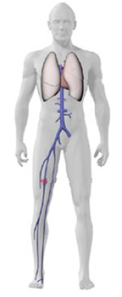
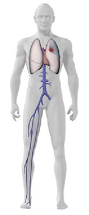
This is when a blood clot known as thrombus develops in the deep veins of primarily the legs and pelvis. It is a condition that requires urgent medical attention to prevent further complications such as a life-threatening pulmonary embolism (PE) or permanent chronic damage to the leg veins, known as post-thrombotic syndrome . It is estimated that approximately 1 in 1,000 people have a DVT each year in the UK. 25,000 deaths per year in England are due to blood clots (PEs that have happened after a DVT) .
SYMPTOMS
Common symptoms include localised pain or tenderness within a calf or thigh muscle, usually associated with ankle and calf swelling.
Blood clots in deep veins can grow in size, break loose, and then travel through the bloodstream to the lungs resulting in life-threatening pulmonary embolism (PE). Symptoms relating to PE include breathlessness, chest pain, palpitations, increased heart rate and coughing.

RISK FACTORS
-
•Risk factors for DVT include
-
•Previous DVT or family history of DVT
-
•Immobility, such as bed rest or sitting for long periods of time
-
•Long journeys by plane, train or coach
-
•Recent surgery
-
•Dehydration
-
•Hormone therapy or oral contraceptives
-
•Pregnancy or post-partum
-
•Previous or current cancer
-
•Limb trauma and/or orthopaedic procedures
-
•Coagulation abnormalities
-
•Obesity
DIAGNOSIS
The most commonly used test to diagnose DVT is an ultrasound scan of the veins in the affected limb, known as a venous duplex scan and a sensitive blood test (D-dimer). MRI Venogram or CT venogram could be used for more detailed visualization of the clot burden, especially in the pelvic area and to plan endovascular treatment. Extremely rarely special x-ray venogram is performed for diagnostic purposes.


Animation showing how a DVT blood clot forms

